
Direct microscopy for the presence of fungal elements & culture for Dermatophyte fungi and other causes of superficial fungal infections.
For the diagnosis of superficial fungal infections of Skin, Hair & Nails.
A clinical diagnosis of fungal nail infection (onychomycosis) can be made without the need for microbiological sampling. https://cks.nice.org.uk/topics/fungal-nail-infection/
Topical agents are recommended as first line therapy. Should this fail, we then recommend sampling to confirm the diagnosis prior to commencing second line (systemic) therapy.
Skin
Hair
Nails
(If you are considering fungal culture from another site/specimen type – please request fungal culture in the clinical details of the culture request for that specimen.)
For electronic requesting using EPIC (RDUH ONLY):
Search for: ‘Mycology MC&S‘ or test code ‘LAB1294‘
Material should be sent in a Dermapak kit (small plastic bag containing black cardboard and instructions – see photo).
This kit is available from pathology stores.
(Please note: The pack is not sterile, so bacterial culture is not appropriate from the same specimen.)
Material from skin lesions is collected by gently scraping off material from the outer edges of the lesion, usually with the edge of a glass microscope slide or a scalpel blade. The edge is most likely to contain viable fungus.
Scalp scrapings are obtained as above but should include hair stubs. Hairs may be plucked from the scalp with forceps, but cut hairs are unsatisfactory as infection is usually below the surface near the scalp. The material should be transported to the laboratory as for skin scrapings.
Clippings should be taken from the discoloured or brittle parts of the nail and cut back as far as possible from the free edge as some fungi are restricted to the lower parts. Scrapings can also be taken from under the nail to supplement the clippings. Nail clippings often fail to grow fungi even if present. (Please note: the laboratory is unable to process whole nails – these specimens will be rejected.)
Additional guidance for specific types of nail infection:
A) Distal subungual infection (The commonest form of infection):
• Clip the distal portion of the nail to expose the abnormal material under the nail plate.
• Using a small curette, dental scraper, blunt scalpel or scissor blade, firmly scrape under the nail plate until the crumbling white degenerating portion is reached, discard the superficial material
• Scrape the keratin debris directly onto a black collection card (Dermapak). This makes it easier to see how much material has been collected
• If the nail is onycholytic it should also be possible to scrape some keratinous material from the underside of the nail clippings
• DO NOT send large pieces of nail clipping
B)Proximal subungual onychomycosis:
Pare down the normal surface of the nail plate at the lunula and collect the white debris from the deeper portion of the plate onto a Dermapak.
C)White superficial onychomycosis:
Scrape the white spots on the nail and discard the outermost surface material; scrape some of the underlying white debris onto a Dermapak.
D)Candida infection:
The material closest to the proximal and lateral nail edges should be obtained. If Candida onycholysis is suspected, the lifted nail bed and, if necessary, the under surface of the nail plate are scraped.
Processed locally in Microbiology at RDUH
Microscopy available within 48 hours. Culture available within 2-3 weeks.
Please note 30-50% of specimens with positive microscopy are negative on culture in any laboratory
Please contact the laboratory prior to sending specimen.
Fungal Tests – Lab Tests Online UK
Fungal Infections – Lab Tests Online UK
Fungal skin infection – scalp | Health topics A to Z | CKS | NICE
Fungal skin infection – body and groin | Health topics A to Z | CKS | NICE
Fungal skin infection – foot | Health topics A to Z | CKS | NICE
Fungal nail infection | Health topics A to Z | CKS | NICE
Specimen Labelling Procedure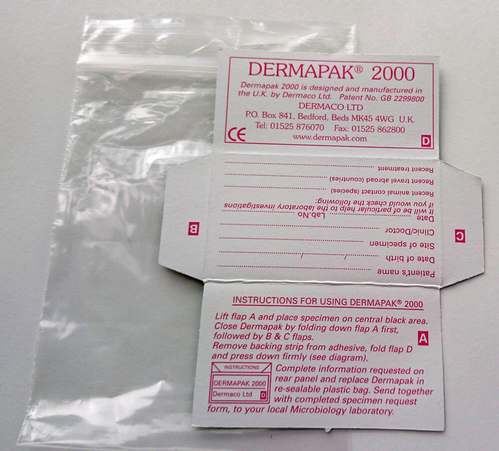


9018
